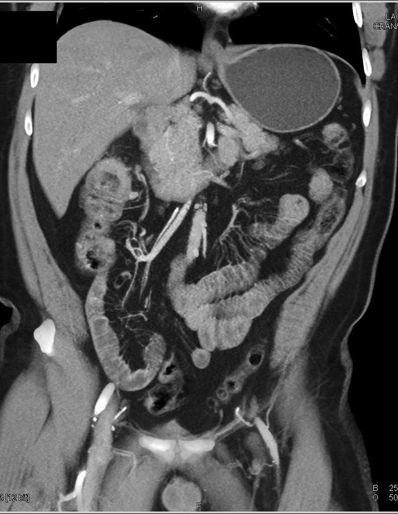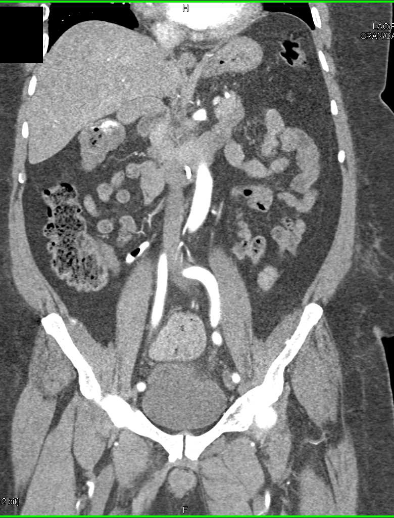
Anatomy figure: 37:06-04 at Human Anatomy Online, SUNY Downstate Medical Center - "The large intestine. 6: Anterior displacement of the DJ flexure with interposition of sigmoid colon, seen in a control patient.(2007) Chapter 4: Abdomen Human anatomy, A clinically-orientated approach.

^ Schneider, Armin Feussner, Hubertus (), Schneider, Armin Feussner, Hubertus (eds.), "Chapter 2 - Anatomy, Physiology, and Selected Pathologies of the Gastrointestinal Tract", Biomedical Engineering in Gastrointestinal Surgery, Academic Press, pp. 11–39, ISBN 978-0-12-803230-5, retrieved.Philadelphia: Elsevier/Churchill Livingstone. Mitchell illustrations by Richard Richardson, Paul (2005). (), "Small Intestine", Imaging Anatomy: Chest, Abdomen, Pelvis (Second Edition), Elsevier, pp. 636–665, ISBN 978-1-9, retrieved
#Ct flexture manuals#
Manuals Plate Girder Shear And Flexural Strengthening Design Example. Introducing Flextur workstations with Gridlok metal pegboard/tool board into that workflow will provide the platform to sort, standardize, and sustain your Lean and 5S efforts. The ligament of Treitz, a peritoneal fold, from the right crus of diaphragm, is an identification point for the duodenojejunal flexure during abdominal surgery. marriott connecticut strawberries and cream hydrangea. Implementing Lean and 5S principles in your organization's workflow increases efficiency, eliminates waste, reduces production time, and decreases lead time. It is covered in front, and partly at the sides, by peritoneum continuous with the left portion of the mesentery. The duodenojejunal flexure lies in front of the left psoas major muscle, the left renal artery, and the left renal vein. : 274 It is retroperitoneal, so is less mobile than the jejunum that comes after it, helping to stabilise the jejunum. The duodenojejunal flexure is surrounded by the suspensory muscle of the duodenum.

At this point, it turns abruptly forward to merge with the jejunum, forming the duodenojejunal flexure. The ascending portion of the duodenum ascends on the left side of the aorta, as far as the level of the upper border of the second lumbar vertebra.


 0 kommentar(er)
